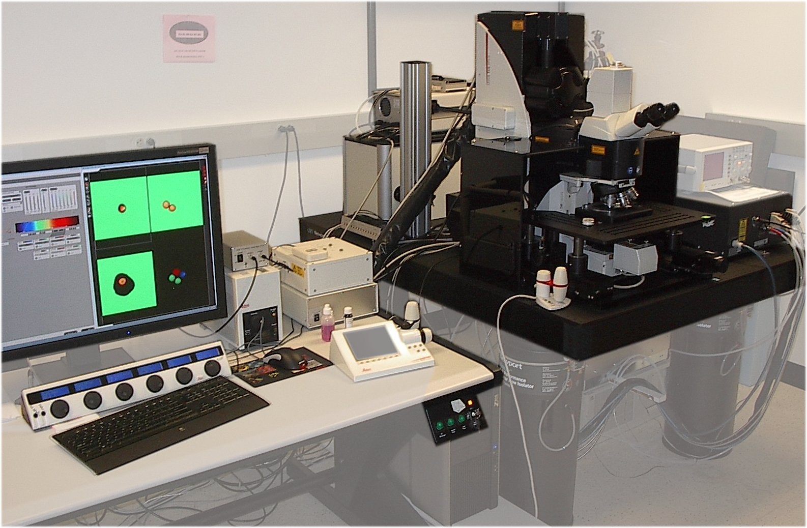
Leica SP5 TCSII with Coherent Chameleon multiphoton (MP) VisionII (IR) laser
TCS (true confocal scanner) SP5 (spectral imaging with up to 5 detectors)
-MP laser tunable from 680nm-1080nm - >3 Watts power at peak output
-MP laser 80MHz, femtosecond pulses make 2PA events likely enough to produce fluorescent micrographs
-also with 405nm and 561nm diode lasers and Argon and HeNe gas lasers with multiple lines selectable (458,476,488,496,514,and633nm)
-with 6 non-descanned detectors, 4 descanned detectors and with TLD (Transmitted Light Detector, PMT) and standard epifluorescence
-with prism/slit based, full spectrum tunable emission filtering
-with AOTF filtering allowing ROI (region of interest) specific illumination
The Leica SP5 is capable of true spectral imaging with up to 5 detectors in the scanhead. MU/MBIC has 4 descanned detectors (DDs) and 6 non-descanned detectors (NDDs).
With Leica SP5 in descanned mode,
the emitted/reflected light passing through the detector pinhole is diffracted by a prism and distributed to four detectors each with a slit allowing mapping of different wavelengths with 1nm spectral resolution. This varying light intensity is transformed into electrical signals by photomultiplier tubes and displayed on the computer monitor screen.
In non-descanned mode (usually MP detection), the detectors are very close to the sample making detection efficiency high. Each non-descanned detector is matched with an emission filter allowing simple spectroscopy with the NDDs as well as the high spectral resolution of the DDs. The NDDs' emission filters cover the visible spectrum. When using NDDs don't forget to turn off the room lights.
There are a number of benefits associated with the use of the Leica SP5 confocal microscope:
• Light rays from outside of the focal plane will not be recorded so unfocused light does not create blurring. Leica LAS/AF(Leica applications suite, advanced fluorescence)software allows conveniently set the pinhole at one Airy unit allowing optimal x/y/z resolution to be obtained.
• Scanning the object in x/y-direction as well as in z-direction allows true, three-dimensional data sets of voxels (volume elements) to be recorded. The MU Leica SP5 has a resonant scanner capable of 16,000 lines per second in x/y plane.
• LAS/AF processiong software enables full 3D reconstruction and analysis.
•Integrated electro-physiology software/hardware allows temporal coordination of imaging with physiological measurements.
•With the Leica SP5 microscope (in non-MP mode) and optimal pinhole, resolution is as low as 200nm in XY and 350nm in z axis.
Further benefits come with Leica SP5 MP micrsocopy:
•In MP mode the z-resolution is further improved (making pinhole unnecessary) due to the superlinear absorption qualities of 2 photon absorption (2PA). Only with very high laser power and very tight focus (high NA lens) will a 2PA event occur; this only happens in a small volume (smaller than 1PA) surrounding the focal 'point' of the lens.
•In MP mode although sample heating (IR induced)can be of some concern, bleaching is much reduced in material surrounding the focal volume.
•In MP mode, penetration depth is increased relative to standard confocal imaging.
SP5 Related links:
• Leica literature relating to the SP5 TCS MP
• objective lenses available for Leica SP5 at MBIC
• information allowing processing of .lif images with NIH imageJ ; from this page, save the loci_tools.jar to your ImageJ plugins folder
Other general confocal links:
The confocal microscope permits examination of living cells without leading to their demise. Using laser light source, confocal microscopy produces sharp fluorescent images because light from only one focal plane is used to produce the image. Light from other "out of focus" planes is blocked by a detector pinhole, part of the optical system of the microscope (see an animation of how the confocal microscope works http://www.olympusconfocal.com/java/confocalsimulator/index.html, an interactive simulation made by Olympus of a similar confocal microscope.
• http://rsb.info.nih.gov/ij/ NIH Imagej free download for image analysis.
•http://www.nephrology.iupui.edu/imaging/software/confocal-assistant.exe Confocal Assistant free download for simple image analysis.
These tools and others are utilized to aid in characterizing and understanding nanomachines, molecular systems and cell biology through research. To discuss use of the system and/or other imaging systems of interest, please contact Dr. Michael L. Norton or refer to the Contact page for further information.


 |
